The formation of α-synuclein amyloid fibrils is a hallmark of the development of Parkinson’s disease. Now researchers have found that α-synuclein can form nanoscale clusters with a very high concentration that can gradually develop into droplets and amyloid fibrils. A researcher says that this may affect how Parkinson’s develops.
Parkinson’s disease is a complex disease that researchers do not yet fully understand. There is therefore currently no cure.
A hallmark of Parkinson’s is the formation of α-synuclein amyloid fibrils in the neurons of the brain. α-Synuclein normally contributes to regulating the flow of information between the brain’s neurons, but when it forms fibrils, this information flow is disrupted, neurons die and disease develops.
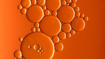
A few years ago, researchers discovered that α-synuclein can form dynamic oil-like droplets that separate themselves from the rest of a protein solution – like droplets of oil in water.
α-Synuclein may be extremely concentrated within these droplets and increase the potential for forming the fibrils associated with Parkinson’s.
Now new research demonstrates how nanoclusters of α-synuclein are formed and develop into droplets and amyloid fibrils and how this knowledge may be important for understanding how Parkinson’s develops.
“If the concentration of α-synuclein is low in a test tube, the fibrils form very slowly. However, the formation of fibrils accelerates rapidly if α-synuclein forms nanoclusters and then droplets. The question is whether this also happens in neurons in the brain and whether the formation of the nanoclusters is the first step towards developing Parkinson’s,” explains a researcher behind the study, Alexander Buell, Professor, Department of Biotechnology and Biomedicine, Technical University of Denmark, Kongens Lyngby.
The research has been published in Nature Chemistry.
Droplets can form in two ways
The researchers discovered that, under some conditions, the α-synuclein droplets formed after a significant delay of a few days.
Other non-amyloidogenic proteins can form similar droplets, but this usually happens very quickly.
The question therefore is whether the droplets in this case form slowly because:
- the droplets do not form easily because this requires much energy and therefore takes time;
- the droplets initially start as nanoclusters that need to grow a few days before they are visible under a microscope.
“It is either one or the other, but we did not know which was more likely,” says Alexander Buell.
Mass photometry revealed the formation of nanoclusters
To answer this question, the researchers from the Technical University of Denmark teamed up with researchers from Novo Nordisk A/S, who had just installed an advanced mass photometry instrument.
Mass photometry can detect and study tiny structures that even an advanced microscope cannot detect.
Alexander Buell says that he estimated that the chance of detecting something when the researchers placed their first sample under the mass photometry instrument was about 5%, but his estimate was way too low.
“The first sample showed that nanoclusters were omni-present. Mass photometry is not sensitive enough to identify all the individual protein molecules but can detect them when they agglomerate to form nanoclusters. Under the microscope we saw something that looked like a swimming pool with many bacteria floating around,” explains Alexander Buell.
Possible new way of forming fibrils
In further research, Alexander Buell and colleagues investigated how the formation of α-synuclein nanoclusters may be related to the formation of fibrils – perhaps also in the brain of people with Parkinson’s.
This showed that the nanoclusters created the ideal conditions for forming fibrils but also that the fibrils could be formed without nanoclusters forming first.
The concentration of α-synuclein, the salt concentration of the liquid and the temperature determine whether one or the other will occur.
“The nanoclusters are an extra aspect in understanding how the fibrils can form. α-Synuclein is one of the most thoroughly studied proteins in the world, so we are very excited that this new study was able to provide new knowledge,” explains Alexander Buell.
He elaborates that researchers within Parkinson’s have known that fibrils can be formed in three very distinct ways and that the new study has now found a fourth way. The question then is whether fibrils are formed in one way or the other or whether several pathways can lead to the development of Parkinson’s.
Other researchers will determine this.
“In addition, this phase separation, which forms droplets of concentrated protein, could be relevant in other diseases such as Alzheimer’s or amyotrophic lateral sclerosis that are also related to protein clumping,” adds Alexander Buell.
New method of studying protein phase separation
In addition to providing new insight into the potential mechanisms behind the formation of fibrils in the brain of people with Parkinson’s, the study also shows that mass photometry can be used to study protein phase separation.
Alexander Buell expects that researchers will jump at this opportunity to study various aspects of phase separation, some of pharmaceutical interest and others in other research fields.
“We have previously lacked opportunities to study protein phase separation at the nanoscale, but we have now shown that mass photometry can accomplish this very easily, which also means that our study has immediately garnered great interest,” he concludes.