Countless studies have shown that exercise is good for the body, but researchers have not had an easy time figuring out why. One reason is that determining what happens inside muscle fibres during exercise training is difficult. A new approach that combines freeze-drying muscle fibres and muscle-proteomic analysis, provides a snapshot of the many changes that occur in muscles with exercise training. The research found thousands of proteins in slow-twitch and fast-twitch muscle fibres and identified how they are regulated with exercise. These new insights may help to better understand diseases and treat the people who have them.
Your pulse increases, sweat trickles down your forehead and you breathe faster. Exercise has an obvious impact on the body. But after just a few workouts, the same effort feels easier because the body adapts. Muscles are key to this adaptation, but seeing and measuring the changes that exercise causes inside the cells while we exercise has been difficult. Muscle biopsies have answered researchers’ questions on oxygenation, substance transport and muscle fibre type for more than 50 years, but really understanding how muscles function and change during exercise training requires delving deeper.
“Previously researchers had several limitations in using muscle biopsies to examine muscle fibre types. The analysis techniques only allowed a few proteins to be analysed due to limited starting material. The other was isolating fibres from fresh and not frozen biopsy material, which was a logistical challenge, since many people have to be ready at the same time: some researchers carry out the biopsies and others stand by to process the biopsies immediately,” explains co-author Jørgen Wojtaszewski, Professor, Molecular Physiology, Department of Nutrition, Exercise and Sports, University of Copenhagen.
Nevertheless, isolating and preparing the muscle fibre under the microscope still takes a long time, and many unwanted changes may have occurred inside the muscle fibre along the way.
"Our new technique enables us to snap-freeze and freeze-dry the samples so that we can carefully separate the small fibres, categorize them by type and analyse how many and which proteins are inside the cells. We were amazed to find many thousands of proteins and the great differences between the fibres and regulation caused by exercise training."
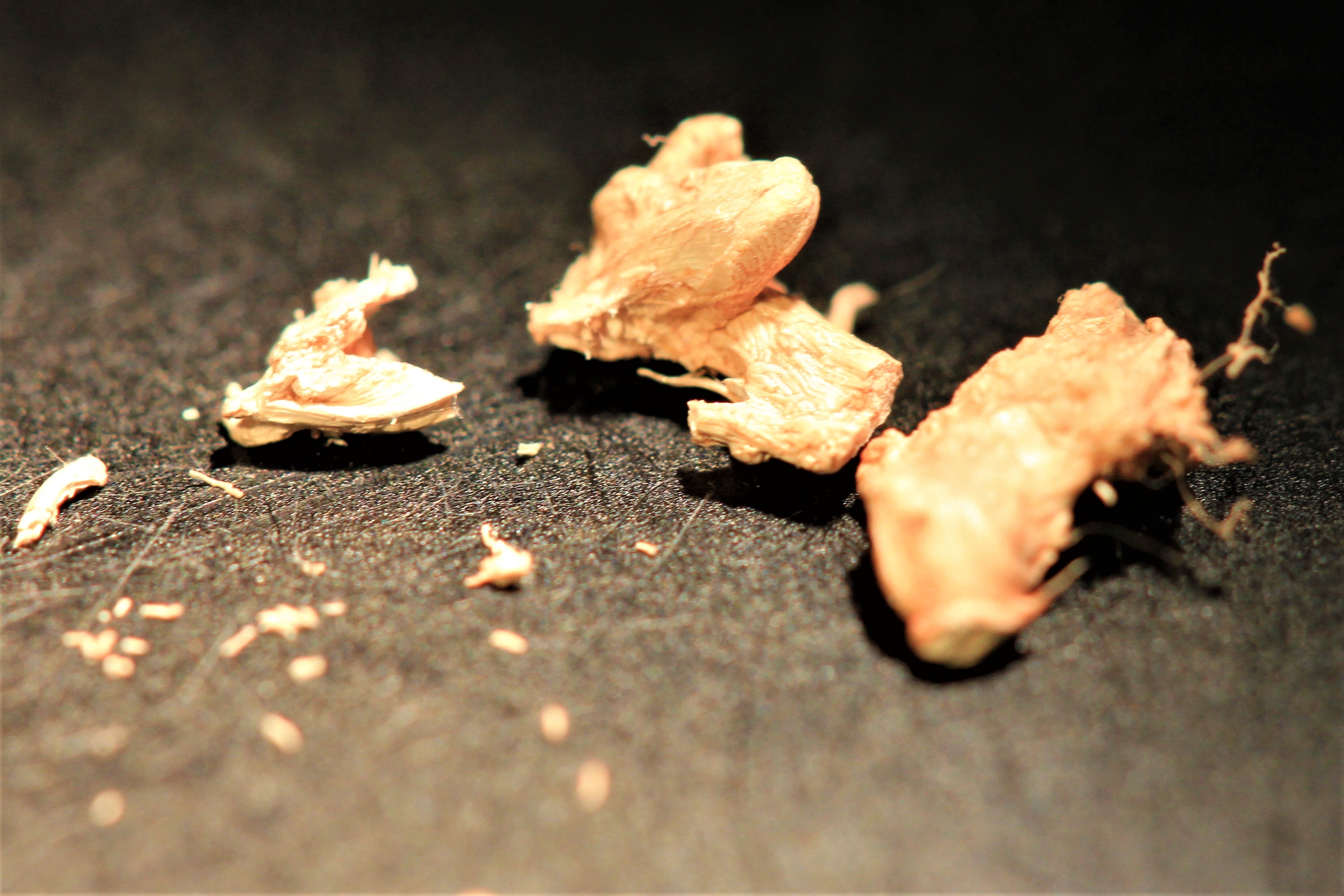
Somewhat like frozen chicken
Exercising regularly is one of the best defences against metabolic disorders, such as obesity and diabetes. To understand why, researchers have tried for years to analyse protein composition and the changes in different types of muscle during exercise.
"Biopsies of muscle tissue usually contain not only muscle fibres but numerous other types of cells," explains co-author Jørgen Wojtaszewski,
Examining the changes specific to the muscle fibres requires that they be isolated from the other cells. In the past, this was done using fresh biopsy material, which is both labour-intensive and renders the fibres unusable for some types of analysis. The new method therefore creates completely new opportunities.
“Immediately after the muscle biopsy is obtained, we freeze the samples in liquid nitrogen so that they can be stored safely in the freezer for a long time. Then we freeze-dry the samples thoroughly. After this, the muscle samples look like a piece of chicken stored in your freezer for too long. In the frozen and dry sample all cellular processes stop. The samples can then be warmed up to room temperature and dissected in a special very dry room so that the samples do not absorb water. Under these conditions we can now separate the muscle fibres individually and make them ready for further analysis,” says Jørgen Wojtaszewski.
More than 4,000 proteins
The researchers used the technique to analyse differences in the slow-twitch muscle fibres that ensure muscle endurance, typically benefitting long-distance runners, and the fast-twitch muscle fibres, typically benefitting sprinters. After separating the muscle fibres, the researchers performed proteomic analysis specific to each type of muscle fibre.
“This enables us to take a snapshot of the muscle cell and measure the expression of thousands of proteins before and after exercise training and thus understand what makes one muscle fibre slow-twitch and another one fast-twitch” explains co-author Atul Deshmukh, Associate Professor, Novo Nordisk Foundation Center for Basic Metabolic Research, University of Copenhagen.
Or illuminate the composition of a normal fibre and compare this to a fibre from ill patients, such as those with diabetes. Surprisingly, they identified more than 4,000 proteins in the samples and showed that exercise training alters the expression of hundreds of proteins in both slow- and fast-twitch muscle fibres.
“This is highly interdisciplinary research involving human physiologists and proteomics experts. Such research was possible due to the state-of-the-art proteomics technologies established at the Novo Nordisk Foundation Center for Protein Research under the leadership of Matthias Mann,” adds Atul Deshmukh.
An incomplete jigsaw puzzle – now with even more pieces
The new study is the most in-depth analysis of slow and fast-twitch muscle fibres ever. It opens up opportunities for future studies but also paves the way for renewed analysis of muscle samples from previous studies located in freezers around the world.
“Our study reveals that this type of analysis creates vast knowledge. The participants’ cycling altered the expression of 237 proteins in the slow-twitch muscle fibres and 172 proteins in the fast-twitch muscle fibres, about 10% of all the proteins identified. We assume that other types of physical activity will alter both the distribution and the number of proteins expressed,” says Atul Deshmukh.
Although the data are a goldmine, the researchers say that finding meaning in the enormous amount of new information is challenging.
“We knew some of this before, and it confirms several theories, but the volume of data is also challenging because the jigsaw puzzle has become much larger and more detailed than before and actually looks more unfinished now, even though we actually know more,” explains Atul Deshmukh.
The jigsaw puzzle may become even more complicated if the researchers get their way. In addition to the different ways the muscle fibres express proteins, the muscle cells also regulate the activity of proteins in different ways, such as being facilitated by small phosphate groups.
“Our next goal is to optimize the analysis to include these post-translational changes such as phosphorylation, so that we not only determine which proteins the muscles express but also how active they are,” says Jørgen Wojtaszewski.
Small changes, great influence
Although this new knowledge is incomplete, it is still very useful. Human skeletal muscles are especially important for understanding the health-promoting effects of exercise, and yet the underlying mechanisms have not been fully elucidated.
“Exercise training alters the physiological behaviour of the muscles, and we are beginning to understand the underlying molecular changes by using the new method. This may turn out to be the missing piece of the puzzle that we need to understand how molecular changes improve our metabolic health,” explains co-author Dorte Steenberg, Postdoctoral Fellow, Molecular Physiology, Department of Nutrition, Exercise and Sports, University of Copenhagen.
Most people have a roughly equal distribution of the two types of muscle fibres, but the ratio can vary greatly between people. This also means that some types of physical activity can benefit certain people more than others.
“Walking may be the healthiest solution for one person, but high-intensity training may be right for someone else with a different distribution of muscle fibres. We know that exercise is even more important for people with type 2 diabetes because it increases insulin sensitivity, and the muscle fibres play a very important role in this."
Clarifying a disease
The new technique enables the researchers to determine whether the problems in the muscles of people with diabetes stem from one or both types of fibres and whether a specific type of exercise could be more beneficial than another.
Measured by weight, skeletal muscles are the largest organ in the human body, and even small changes can strongly affect metabolism in the whole body. Skeletal muscles are therefore interesting from a pharmaceutical perspective, since they have great potential in the treatment of people with metabolic diseases.
One challenge, however, is to avoid side-effects in heart muscle, which comprises specialized fibres similar to slow-twitch skeletal muscle fibres.
“In theory, if we can identify proteins that the fast-twitch muscle fibres specifically express, we may be able to guide drugs into precisely these fibres – and in this way perhaps avoid side-effects in the heart muscle. Some muscle disorders primarily affect one type of muscle fibre: for example, the fast-twitch fibres are those primarily affected in Duchenne muscular dystrophy. Our method could be used to further clarify this disease and perhaps lead to therapy precisely targeting these muscle fibres,” concludes Jørgen Wojtaszewski.