When people digest food, the flow of fluids and digestive enzymes to the gut is critical. Until recently, researchers had marvelled at the incredibly complex system of ducts that transports a stream of enzymes and mucus from the pancreas to the gut. Astonishingly, a Danish research project has revealed that the ducts are created similar to river beds. This new knowledge may lead to improving treatment for people with cystic fibrosis and diabetes.
Most people associate transport with roads, rivers or rails. In nature, transport is equally vital. Trees have a transport system in their roots, branches and leaves, and humans have many transport channels such as nerve fibres and lung bronchioles. Many of these structures are nearly identical from person to person. However, this does not apply to the transport ducts between the pancreas and the gut, which are essential for efficient digestion. A Danish research project has now solved the enigma of how the ducts are created to transport mucus, enzymes and chemical substances.
“The transport channels from the pancreas to the gut are critical for digesting food and neutralizing acidic gastric juices. Because efficient transport is essential, we wondered why the ducts vary from person to person. Our new results show that the channels can change similarly to a river bed during fetal life. The channels with the greatest flow appear to widen, whereas others run dry and disappear. This knowledge may help us to treat people with cystic fibrosis and some forms of monogenic diabetes associated with cystic ducts, who have such transport problems,” explains Anne Grapin-Botton, Professor, Novo Nordisk Foundation Center for Stem Cell Biology, University of Copenhagen.
A surprising result
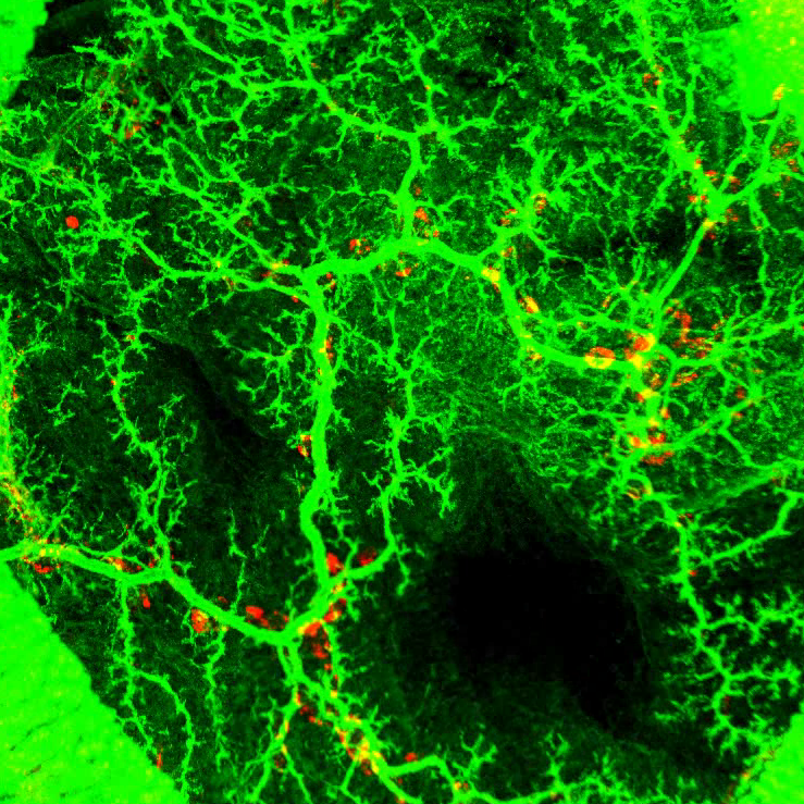
To map how the ducts inside the pancreas are formed, the researchers marked the ducts with fluorescent antibodies, which enabled them to see when a duct was formed and its connection to others. The researchers monitored the development of the pancreas in mice.
“During organ development in fetuses, the ducts are initiated from small holes. These small holes connect and fuse together, thereby creating many ducts that develop into a complex network. It resembles a town with a labyrinth of streets. What fascinated us was how, from this labyrinth, a simpler treelike structure emerged at birth.”
To keep track of the high volume of data and to identify how the network of ducts changed, the researchers teamed-up with Kim Sneppen, a professor and physicist from the Niels Bohr Institute, University of Copenhagen. Svend Bertel Dahl-Jensen, their PhD student, pulled together the threads and uncovered the secrets of the labyrinth.
“We decided to adapt the programs used to model road, rail or internet networks, and the picture that emerged once we inputted the data into the computer reminded us of something familiar: a river system. And indeed, going back to the laboratory, we found that as soon as the ducts were formed they secreted juice. We think that this creates a flow of fluid towards the intestine already in fetuses. Some ducts widen, while others run dry and disappear.”
Ducts collapse
The study showed that the cells from the collapsed ducts did not simply disappear. They are likely recycled to widen existing ducts. This may turn out to be important for medicine.
“People with cystic fibrosis or with certain forms of monogenic diabetes have problems in the pancreatic ducts. For diabetes, we do not know why diabetes and enlarged ducts are associated when certain genes are mutated.”
People with cystic fibrosis have defective pancreatic ducts. This results from mutations in a gene that codes for a channel enabling fluid secretion inside the ducts. The researchers demonstrated that the secretion defects may start very early in fetuses and their work may lead them to consider earlier treatment.
“We would now really like to understand why people whose pancreatic ducts have collapsed are more likely to develop diabetes. One hypothesis we are pursuing is that they make less beta cells secreting insulin. These cells are initially formed in the ducts. We are now studying this in mouse models and in miniature human pancreas (organoids) made from stem cells in 3D culture.”
“Deconstructing the principles of ductal network formation in the pancreas” has been published in PLOS Biology in collaboration between DanStem and the Niels Bohr Institute of the University of Copenhagen, supported by the Danish National Research Foundation. Anne Grapin-Botton is a professor affiliated with the Novo Nordisk Foundation Center for Stem Cell Biology of the University of Copenhagen. The Novo Nordisk Foundation awarded research grants of almost DKK 700 million to the Center in 2010–2017.